吉林大学-Food & Function:红景天苷通过调节肠道微生物群缓解葡聚糖硫酸钠诱导的小鼠结肠炎
吉林大学临床兽医学系曹永国教授团队在Food & Function发表题目为“Salidroside alleviates dextran sulfate sodium-induced colitis in mice by modulating the gut microbiota”研究论文(红景天苷通过调节肠道微生物群缓解葡聚糖硫酸钠诱导的小鼠结肠炎)。
摘要:
菌群失调导致炎症性肠病(IBD)持续进展。在本研究中,我们旨在探索从红景天中提取的主要糖苷红景天苷(Salidroside,Sal)是否可以通过调节微生物群改善葡聚糖硫酸钠(DSS)诱导的结肠炎。结果表明,口服15 mg kg-1的Sal可抑制DSS诱导的小鼠结肠炎,表现在结肠长度、组织学分析、疾病活动指数(DAI)评分以及肠道中巨噬细胞的比例和数量。Sal还部分恢复了结肠炎小鼠的肠道微生物群。为了验证因果关系,设计了一项粪菌移植(FMT)研究。与DSS处理的小鼠相比,Sal处理的供体小鼠的FM显著减轻了结肠炎小鼠的症状,包括降低DAI评分,减轻组织损伤,促进黏蛋白( mucin-2)和紧密连接(TJ)蛋白(occludin和闭锁小带-1 (ZO-1))的表达,减少肠道中的M1型巨噬细胞。结果发现,Sal和FMT均影响了肠道菌群的结构和丰度,在属水平上表现为Turicibacter、Alistipes、Romboutsia相对丰度的降低和Lactobacillus相对丰度的增加。此外,当用抗生素耗竭肠道微生物群时,Sal的抗炎作用消失,证明Sal以肠道微生物群依赖的方式减轻肠道炎症。因此,Sal可以作为结肠炎功能性食品的显著候选者。
研究结果:
Sal给药改善了DSS诱导的急性结肠炎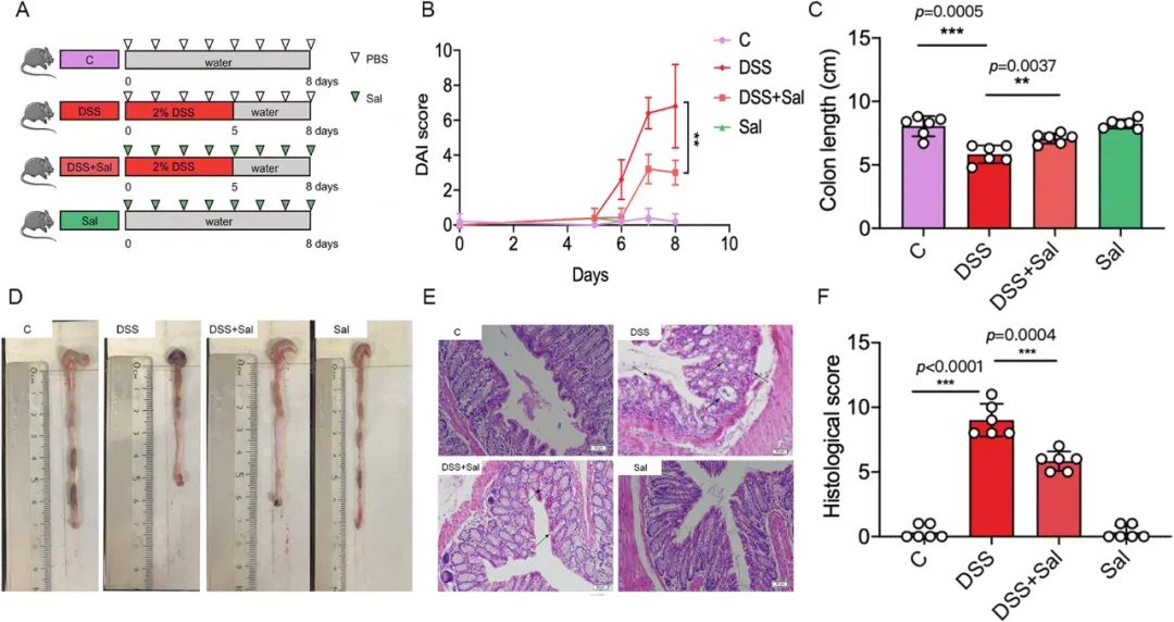
Fig. 1 Sal administration alleviated DSS-induced colitis in mice. (A) Study design – the mice were given 2% DSS in drinking water for 5 days and then provided with water for another 3 days before being sacrificed. The Sal was given daily by oral gavage (15 mg kg−1). (B) The DAI score was measured. (C) and (D) The colon length of the mice was determined (c), and the representative colon images are shown in (D). (E) Representative images of the H&E staining, scale bar: 50 μm. (F) The histological score of the H&E staining was determined. Data are means ± SD of at least three independent experiments (n = 6). *p < 0.05, **p < 0.01, ***p < 0.001.
Sal给药减少了结肠炎小鼠中的促炎巨噬细胞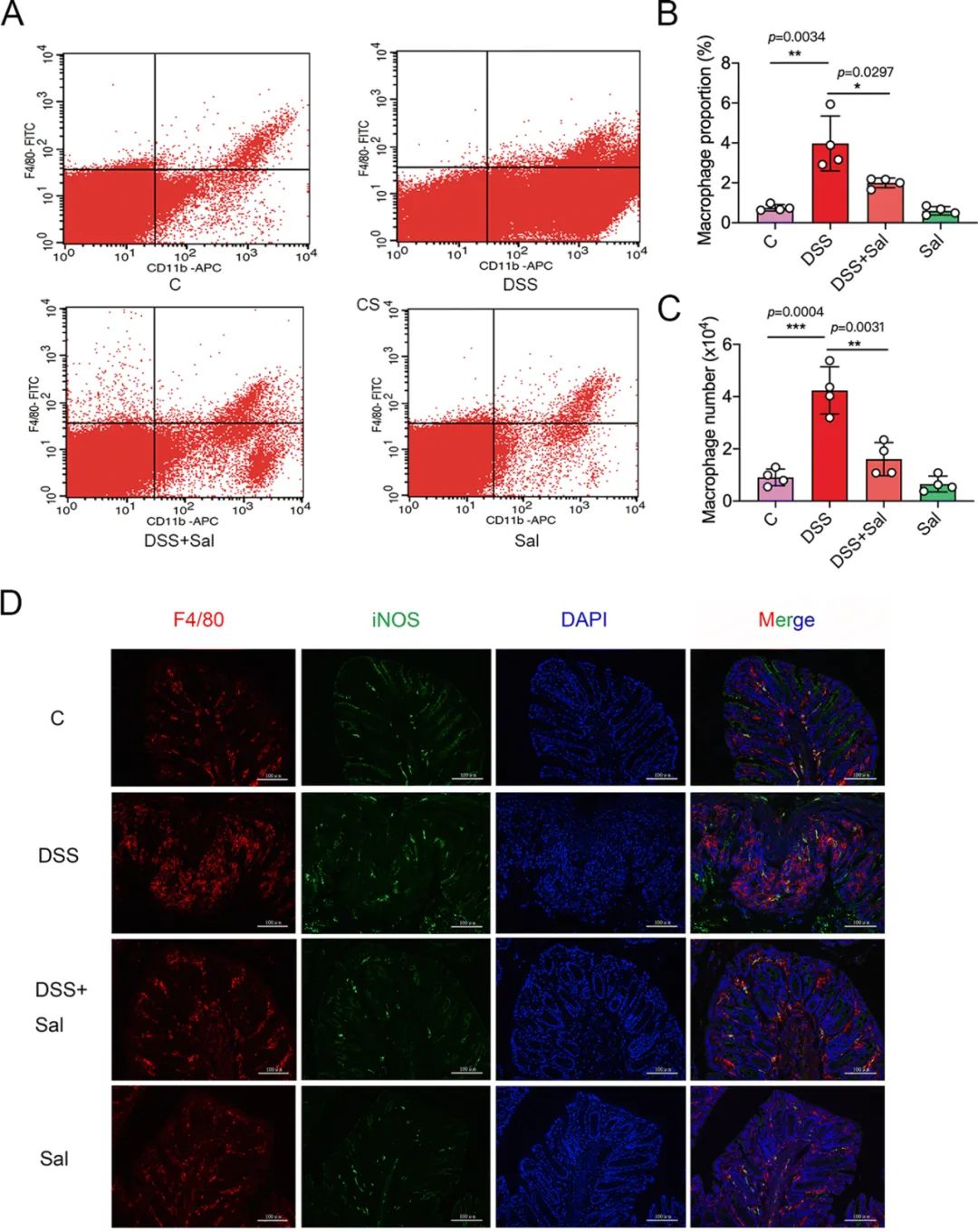
Fig. 2 Sal administration reduced pro-inflammatory macrophages in the intestine of colitic mice. (A) Representative image of flow cytometry. The proportion (B) and the number (C) of macrophages in the intestine were determined. (D) IF analysis of f4/80 and iNOS in the mouse colons. Scale bar, 100 μm (n = 4). *p < 0.05, **p < 0.01, ***p < 0.001.
Sal给药调节了结肠炎小鼠的肠道菌群失调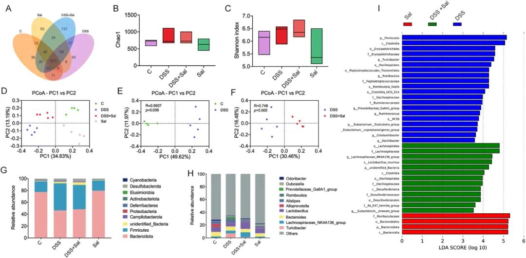
Fig. 3 The effects of Sal administration on gut microbiota in mice with DSS-induced colitis. (A) Venn diagram showing the overlap of the OTUs identified in the gut microbiota among the four groups. The Chao1 (B) and Shannon indices (C) of the fecal microbiota. (D)–(F) The principal coordinates analysis (PCOA) plot of the gut microbiota based on the Bray–Curtis metric. (G) The top 10 microbiota taxa found at the phylum level. (H) The top 10 microbiota taxa found at the genus level. (I) The distribution histogram based on LDA (LDA > 3.5) (n = 4–5).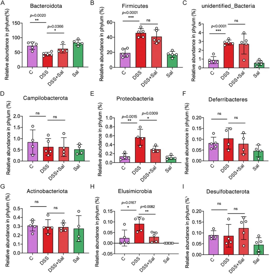
Fig. 4 The profiles of gut microbiota at the phylum level in the four groups. (A)–(I) The selected bacteria at the phylum level (n = 4–5). *p < 0.05, **p < 0.01, ***p < 0.001, ns: not significant.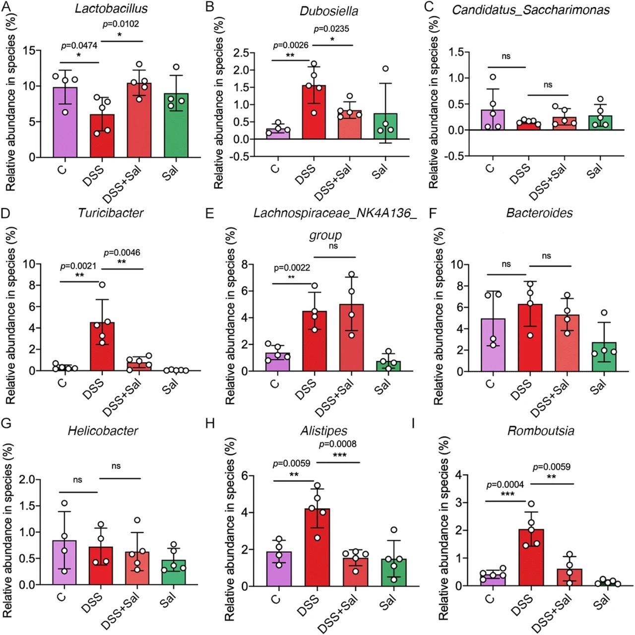
Fig. 5 The profiles of gut microbiota at the genus level in the four groups. (A)–(I) the selected bacteria at the genus level (n = 4–5). *p < 0.05, **p < 0.01, ***p < 0.001, ns: not significant.
Sal处理的小鼠粪便微生物群恢复了结肠炎相关的肠道损伤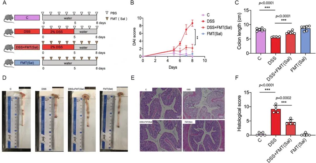
Fig. 6 The Sal-treated FMT alleviated the severity of DSS-induced UC in mice. (A) Study design. Mice were given 2% DSS in drinking water for 5 days and then provided with water for another 3 days before being sacrificed. Mice of the DSS + FMT (Sal) group and FMT + (Sal) group were given 200 μl of fecal microbial suspensions, by oral gavage, from mice of the Sal group for 8 days on a daily basis. The mice of the control group and the DSS group were given 200 μl of PBS by oral gavage. (B) The DAI score was measured. The colon length of the mice was determined (C), and the representative colon images are shown in (D). (E) Representative images of H&E staining (magnification of 100×). (F) The histological score of H&E staining was determined, and the data are means ± SD of at least three independent experiments (n = 6). **p < 0.01. ***p < 0.001.
Fig. 7 The Sal-treated FMT promoted the expression of mucin-2 in colitic mice. (A) Alcian blue analysis of mucin-2 in colons (magnification of 100×). (B) The IF analysis of mucin-2 in colons (magnification of 100×). (C) The mucin-2 mean fluorescence intensity per crypt, data are means ± SD of at least three independent experiments (n = 6). **p < 0.01, ***p < 0.001.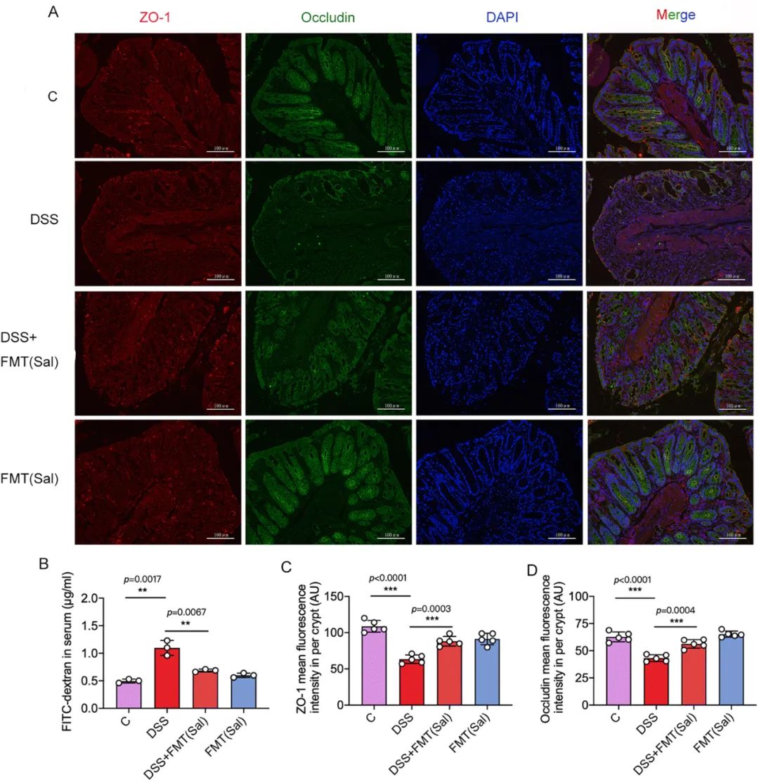
Fig. 8 The Sal-treated FMT improved the intestinal permeability in colitic mice. (A) The IF analysis of ZO-1 and occludin in colons, scale bar: 100 μm. (B) The serum concentrations of FITC–dextran. (C) and (D) The mean fluorescence intensity per crypt for ZO-1 and occludin, data are means ± SD of at least three independent experiments (n = 3–5). **p < 0.01, ***p < 0.001.
Sal对巨噬细胞亚型的有益作用可通过FMT传递
Fig. 9 Sal-treated FMT reduced pro-inflammatory macrophages in the intestines of colitic mice. (A) A representative image of flow cytometry. The proportion (B) and the number (C) of macrophages in the intestine were determined. (D) The IF analysis of f4/80 and iNOS in colons, scale bar: 100 μm. The data are means ± SD of at least three independent experiments (n = 3–4). *p < 0.05, **p < 0.01.
结论:
总之,我们的研究证明,Sal给药通过调节肠道菌群失调显著缓解UC。FMT和肠道菌群耗竭研究旨在验证因果关系。我们发现,Sal和FMT均影响了肠道菌群的结构和丰度,表现为在属水平上,Turicibacter、Alistipes、Romboutsia的相对丰度降低,Lactobacillus的相对丰度增加。因此,我们的研究结果表明,Sal具有作为功能性食品或UC治疗新策略的潜力。
座机
021-58390070
发送您的留言
微信扫码咨询

